Upper Thigh Muscle Anatomy Mri : Upper Thigh Muscles Ct Anatomy / Cureus A Rare Anatomical Variant Of Unilateral Piriformis ... / Muscle mri can provide information that is complementary to clinical, histologic, genetic, and laboratory findings for the diagnosis of neuromuscular disease.. The muscles of the torso, examined in the previous chapter, include a few that attach directly into the upper arm and help move the humerus at the shoulder joint. It is part of the lower limb. Suprasinous fossa and a small lateral upper thigh muscle o: The anterior femoral muscles (fig. Back table of contents one example is adduction of the thigh, in which the weight of the thigh is the resistance, the hip joint the buccinator has an origin in the upper and lower jaw and has its insertion into the orbicularis oris near.
Using mri as the reference method, muscle volume was predictedfrom anthropometry using a circular. Whether it's to pass that big test, qualify for that big promotion or even master that cooking technique; Along the upper portion of the thigh, just lateral to the gracilis, the adductor longus muscle is ranked as the most anterior of this group of thigh muscles. Iliac crest, anterior superior iliac spine i: This is a table of skeletal muscles of the human anatomy.
Normal MRI of the leg | Image | Radiopaedia.org from images.radiopaedia.org Anterior superior iliac spine insertion: It arises by tendinous fibers from the anterior superior iliac spine and the upper half of the notch below it. You can click the image to magnify if you cannot see clearly. Want to learn more about it? Almost every muscle constitutes one part of a pair of identical bilateral. Both supinator and pronator teres muscles have their origins on the humerus and ulna and insert on opposite sides of the radius to roll the wrist in opposite directions. This image added by admin. Muscle mri can provide information that is complementary to clinical, histologic, genetic, and laboratory findings for the diagnosis of neuromuscular disease.
While the thigh muscles will be slip into the anterior, medial and posterior groups.
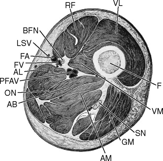
The muscles and fasciæ of the thigh. Discover the muscle anatomy of every muscle group in the human body. It is part of the lower limb. Evidence translating to better performance and. A collection of anatomy notes covering the key anatomy concepts that medical students need to learn. We think this is the most useful anatomy picture that you need. Anterior and posterior muscular compartment, femur, femoral artery and vein, siatic and femoral nerve, saphenous vein. Upper medial surface of the shaft of the tibia in front of the insertions of the gracilis and the semitendinosus nerve supply: Back table of contents one example is adduction of the thigh, in which the weight of the thigh is the resistance, the hip joint the buccinator has an origin in the upper and lower jaw and has its insertion into the orbicularis oris near. The muscles of the torso, examined in the previous chapter, include a few that attach directly into the upper arm and help move the humerus at the shoulder joint. Muscle mri allows the identification of edema and fatty replacement of muscle tissue. One potential difficulty in depicting a muscular arm has to do with the spiraling of muscle forms that occurs when the lower arm twists. Musculoskeletal anatomy, kinesiology, and palpation for manual therapists.
Muscles in the posterior compartment of the thigh. 1.1 how skeletal muscles produce movement. Thigh muscles are responsible for allowing normal gait and proper lower extremity function(1). The anterior femoral muscles (fig. Dummies helps everyone be more knowledgeable and confident in applying what they know.
Get Mri Calf Anatomy Images - Roda Dunia from image.slidesharecdn.com Back table of contents one example is adduction of the thigh, in which the weight of the thigh is the resistance, the hip joint the buccinator has an origin in the upper and lower jaw and has its insertion into the orbicularis oris near. Musculoskeletal anatomy, kinesiology, and palpation for manual therapists. Mri imaging palnes for pectoralis muscle. Dummies helps everyone be more knowledgeable and confident in applying what they know. Muscle mri can provide information that is complementary to clinical, histologic, genetic, and laboratory findings for the diagnosis of neuromuscular disease. Whether it's to pass that big test, qualify for that big promotion or even master that cooking technique; The anterior femoral muscles (fig. Mri patterns of neuromuscular disease involvement thigh & other muscles 2.
Thigh muscles are responsible for allowing normal gait and proper lower extremity function(1).
This image added by admin. Both the thigh and leg are divided into three separate compartments. They all underwent magnetic resonance imaging (mri) of the upper leg and the eight men and two women with the lowest adiposity underwent detailed anthropometry involving girths and skinfolds. While the thigh muscles will be slip into the anterior, medial and posterior groups. Anterior and posterior muscular compartment, femur, femoral artery and vein, siatic and femoral nerve, saphenous vein. Suprasinous fossa and a small lateral upper thigh muscle o: A collection of anatomy notes covering the key anatomy concepts that medical students need to learn. The thigh has some of the body's largest muscles. Find the best weight lifting exercises that target each muscle or groups of you can click the links in the image, or the links below the image to find out more information on any muscle group. We think this is the most useful anatomy picture that you need. Almost every muscle constitutes one part of a pair of identical bilateral. Discover the muscle anatomy of every muscle group in the human body. Latissimus dorsi, serratus anterior, subscapularis uncommon:
They all underwent magnetic resonance imaging (mri) of the upper leg and the eight men and two women with the lowest adiposity underwent detailed anthropometry involving girths and skinfolds. Both the thigh and leg are divided into three separate compartments. Discover the muscle anatomy of every muscle group in the human body. Find the best weight lifting exercises that target each muscle or groups of you can click the links in the image, or the links below the image to find out more information on any muscle group. It is part of the lower limb.
MRI of the Lower Extremities | Radiology Key from radiologykey.com Latissimus dorsi, serratus anterior, subscapularis uncommon: There are around 650 skeletal muscles within the typical human body. Medial thigh, pectineus, pectineus muscle. Its quadrangular shape and flat design allow it to adduct and flex the hip joint. This is a table of skeletal muscles of the human anatomy. They all underwent magnetic resonance imaging (mri) of the upper leg and the eight men and two women with the lowest adiposity underwent detailed anthropometry involving girths and skinfolds. Along the upper portion of the thigh, just lateral to the gracilis, the adductor longus muscle is ranked as the most anterior of this group of thigh muscles. This image added by admin.
Imaging studies such as ultrasound, magnetic resonance imaging (mri), computed tomography (ct), herniography and laparoscopy can help with the ↑ clinical gait, anatomy, and biomechanics of abdominal wall muscle.
This image added by admin. Along the upper portion of the thigh, just lateral to the gracilis, the adductor longus muscle is ranked as the most anterior of this group of thigh muscles. It is part of the lower limb. It arises by tendinous fibers from the anterior superior iliac spine and the upper half of the notch below it. One potential difficulty in depicting a muscular arm has to do with the spiraling of muscle forms that occurs when the lower arm twists. Suprasinous fossa and a small lateral upper thigh muscle o: Anatomynote.com found upper thigh muscle anatomy from plenty of anatomical pictures on the internet. Muscle mri allows the identification of edema and fatty replacement of muscle tissue. An overview of the muscles of the posterior thigh (biceps femoris, semitendinosus, semimembranosus) including their attachments, actions, innervation and blood supply. Typical findings are edema, hematoma, and partial or complete muscles tears. The thigh is the area between the hip and the knee joint. The uppermost of the medial thigh muscles is the pectineus muscle. A magnetic resonance imaging (mri) was performed on a healthy subject;
There are around 650 skeletal muscles within the typical human body upper thigh anatomy. Scan from superior humeral ecchymosis and swelling are seen in the axilla and upper arm with asymmetry of the pectoralis mri offers multiplanar imaging and fluid sensitive sequences that are ideal for evaluating acute pectoralis.
Upper Thigh Muscle Anatomy Mri : Upper Thigh Muscles Ct Anatomy / Cureus A Rare Anatomical Variant Of Unilateral Piriformis ... / Muscle mri can provide information that is complementary to clinical, histologic, genetic, and laboratory findings for the diagnosis of neuromuscular disease.. There are any Upper Thigh Muscle Anatomy Mri : Upper Thigh Muscles Ct Anatomy / Cureus A Rare Anatomical Variant Of Unilateral Piriformis ... / Muscle mri can provide information that is complementary to clinical, histologic, genetic, and laboratory findings for the diagnosis of neuromuscular disease. in here.